Stroke
How are strokes diagnosed?
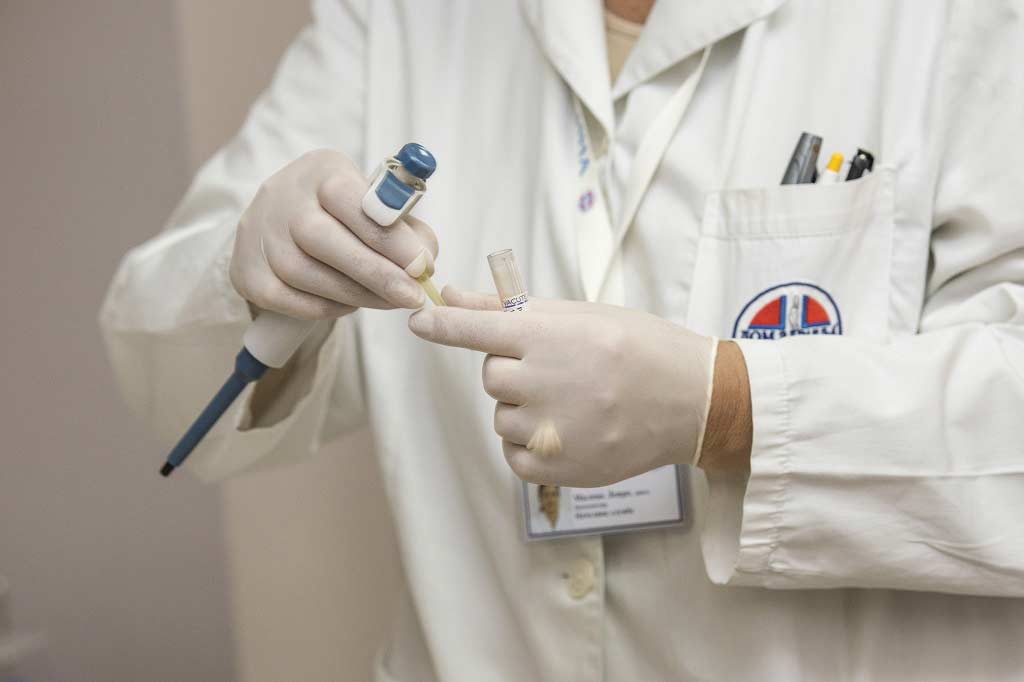 Typical Symptoms Shortly after the patient arrives at the hospital, physicians look for the typical symptoms of a stroke. These symptoms involve Symptoms evident in the face, its retraction to one side (the face seems to droop on one side) Problems with the limbs, (usually arms), hands and legs are numb or cannot be moved Problems with speech, and time during which these symptoms have occurred. A history of the condition is compiled with the help of the patient, if they are capable or from family members. Following these, a series of preliminary blood tests are ordered: A blood test for lipid (fats) levels (a lipid fraction) Checking the heart rate for any heart rhythm disorders Measurement of arterial pressure Following these tests, visual examinations are performed Brain CT-Scan If the symptoms the patient is exhibiting are not indubitably those of a stroke, then a brain CT-scan can indicate: Whether a hemorrhagic insult has occurred, meaning has there been rupture of a blood vessel, or rather an ischemic insult, meaning the obstruction of a blood Which part of the brain has been affected How serious the stroke is Any patient which is experiencing stroke or stroke-like symptoms, should see a doctor within an hour of experiencing their symptoms. They should immediately take a brain scan in order to either eliminate or confirm the diagnosis of a stroke. A contrast scan is a three-dimensional X-Ray which takes extremely thin cross-sections. The patient is intravenously injected with a special liquid called contrast, which allows for doctors to view the blood vessel pathways of your entire brain in great detail. This then allows your physician to clearly see exactly what has occurred, whether the stroke is hemorrhagic or ischemic. A scan is an excellent way to receive precise information about this condition quickly, in order for physicians to immediately decide on a choice of treatment. Magnetic Resonance Imaging of the Brain (Brain MRI) An MRI employs a powerful magnetic field and radio waves in order to produce detailed views of the body, in this particular case of the brain. An MRI is necessary in order to receive more important details related to a certain condition in the brain, it can help localize and relay information about very small areas in the brain. These areas may be involved in the damages caused by a stroke. The Swallow Test In case a stroke has occurred, it is important for the doctor to determine whether or not the patient has retained their ability to swallow. This is of a vital importance, since if the patient has lost their ability to swallow, the foods or liquids they may try to ingest may pass through the respiratory tract and cause serious conditions such as aspiration pneumonia. The test for this is very simple, the patient is asked to drink a sip of water, or other liquid, and if they can swallow without coughing, then the doctor asks them to drink an entire glass of water. If problems are encountered, then a special therapist for speech and the tongue is recruited. When the patient has trouble swallowing, then nourishment is provided intravenously or through a tube introduced from the nose to the stomach (nasogastric tube). Heart and Blood Vessel Tests In order to determine the causes of strokes several blood vessel tests are used. The carotid arteries are scanned, in order to observe either their narrowing or obstruction which may have caused the stroke. If deemed necessary this examination should be conducted within 48 hours. An Echocardiogram A heart echocardiogram is an examination which reveals the morphological and hemodynamic parameters of the heart. An echocardiogram can also be performed through the esophagus, via a probe inserted through the mouth into the esophagus, reaching behind the heart. This is also called a transesophageal echocardiogram. Through this examination one can view the heart and identify any blood clots that may have caused the stroke or any other anomalies.
Introduction
A stroke is a serious and life-threatening medical condition that occurs when the blood supply to part of the brain is cut off. Strokes are a medical emergency and urgent treatment is essential because the sooner a person receives treatment for a stroke, the less damage is likely to happen.
Symptoms and signs of stroke
If you suspect that you or someone else is having a stroke, phone 999 immediately and ask for an ambulance. Even if the symptoms of a stroke disappear while you are waiting for the ambulance to arrive, you or the person having the stroke should still go to hospital for an assessment.
Causes of stroke
There are two main types of stroke - ischaemic strokes and haemorrhagic strokes - which affect the brain in different ways and can have different causes. Ischaemic strokes are the most common type of stroke.
Diagnosing stroke
Strokes are usually diagnosed by carrying out physical tests and studying images of the brain produced during a scan. Even if the physical symptoms of a stroke are obvious, brainscans should also be carried out to determine: if the stroke has been caused by a blocked artery (ischaemic stroke) or burst blood vessel (haemorrhagic stroke), which part of the brain has been affected, how severe the stroke is.
Treating stroke
Effective treatment of stroke can prevent long-term disability and save lives. If your blood pressure is too high, you may be offered medicines to lower it. Medicines that are commonly used include: thiazide diuretics, angiotensin-converting enzyme (ACE) inhibitors, calcium channel blockers, beta-blockers, alpha-blockers. Some ischaemic strokes are caused by narrowing of an artery in the neck called the carotid artery, which carries blood to the brain. The narrowing, known as carotid stenosis, is caused by a build-up of fatty plaques.
Recovering after a stroke
Two of the mostcommon psychological problems that can affect people after a stroke are: Depression many people experience intense bouts of crying and feel hopeless and withdrawn from social activities, anxiety where people experience general feelings of fear and anxiety, sometimes punctuated by intense, uncontrolled feelings of anxiety (anxiety attacks). The injury to the brain caused by a stroke can lead to widespread and long-lasting problems.
Preventing strokes
The best way to prevent a stroke is to eat a healthy diet, exercise regularly, and avoid smoking and excessive consumption of alcohol. Ensuring a balance in your diet is important. If you have not fully recovered from your stroke, you may find that you will have become particularly sensitive to alcohol and even the recommended safe limits as above for the general population may be too much for you.
Patient story: "We call ourselves stroke survivors, not patients - that's very important"
Jim Whyte was forced to give up work after having a stroke, but he's proved that there is life after a stroke. Jim had high blood pressure and was diabetic, which are both risk factors for stroke. However, he had never smoked and, due to his diabetes, was already following the healthy diet recommended for stroke survivors.
Patient story: "Everyday was a new challenge."
Having a stroke on the first day of his summer holiday was the last thing Stephen Harnet expected, especially as he was a healthy 32-year-old at the time. It turned out that the stroke was due to a condition I was born with called AVM (arteriovenous malformation), which is a tangle of abnormal blood vessels (arteries and veins), and can affect the brain and lead to a stroke. Before the stroke I was a technical manager working 12-hour days, seven days a week.
Patient story: "I'm sure the doctors thought I wouldn't get better, let alone run a marathon."
David Diston had a major stroke that left him paralysed down his right side and unable to speak. Now he has made a near total recovery, and has even run a marathon. He woke up in an assessment ward at Swindons Princess Margaret Hospital. He had no feeling or movement down his right side, and he couldn't speak. After a few days, he was moved to a specialist stroke unit where the doctors explained that he would have to learn to speak, write and walk again from scratch.
Patient story: "I was just 26 and had my whole life ahead of me."
Manjit Bains was just 26 when he had a stroke. Determination, support from his family and friends and rehabilitation helped him get his life. "This hasn't been a solo journey. I've had a lot of support, including psychologists and counselling. After intensive care I was nursed on a medical ward at the hospital where I had been working."
Patient story: “Doctors said I had had a massive stroke.â€ÂÂ
Before June/ 17/ 2000, Jane Stokes CBE was a successful lawyer with a 25-year career in the civil service. She says her lifestyle may have raised her risk of a stroke. As a smoker, she had developed a two-pack-a-day habit. She drank moderately and neglected her fitness when her career began to take over. Recovery is a long process.
What is a stroke?
A stroke is a severe condition and a serious threat, which requires immediate medical intervention. This disease is a major cause for invalidity and mortality in the world. Strokes are ranked third in the world in regards to mortality.
Is a stroke a disease that develops gradually, or does it occur suddenly?
Strokes occur suddenly and all at once; they pose a very serious threat to a patient’s life. This condition requires immediate medical attention.
Can you die from a stroke?
A stroke is a very severe condition. Immediate medical attention is necessary; the medical staff usually conducts a series of examinations and medical procedures to offer first aid.
How does a stroke occur?
Due to another disease or condition, a blood vessel in a certain area of the brain may rupture (break) and cause the blood to spread to the surrounding tissue and damage it.
What are the main symptoms of strokes?
At the onset of a stroke, a few main symptoms should be notices, which are easy to remember by using the acronym F.A.S.T. (Face, Arms, Speech, Time).
What is the difference between a heart attack and a stroke?
A myocardial infarction (MI), commonly known as a heart attack, occurs when a portion of the heart is deprived of oxygen due to blockage of a coronary artery. A stroke is a medical condition medical condition that occurs when the blood supply to part of the brain is cut off.
Can stress and anxiety cause a stroke?
A person’s emotional state is an important factor in the function and development of their daily activities. Anxiety, depression and stress are conditions which compromise and affect a person’s daily life.
Which are the major risk factors for a stroke?
Risk factors for stroke, that are worth mentioning include: Atherosclerosis (Depositing of lipids in the lumen of blood vessels in the form of plaques), High blood pressure, Lack of physical activity, Obesity, Continuous consumption of alcohol, Smoking, etc.
What are some of the warning signs of a stroke?
It could occur that prior to a patient experiencing a stroke, they may experience warning signs. A few days or weeks in advance they may experience light symptoms such as unusual headaches, a light lip distortion, numbness in the arm or leg, vertigo, brief loss of vision, difficulties articulating, amnesia, etc.
What types of stroke are there?
There are different types of Stroke, they include: brain stroke of the ischemic type, brain stroke due to hemorrhage (cerebral hemorrhage) and hemorrhages due to the rupture of blood vessels or due to vascular malformation, etc.
What is the cause of a stroke?
A stroke due to a hemorrhage occurs more rarely than a cerebral ischemia. The major cause for this type of stroke is arterial hypertension.
What are the causes of TIA (Transitory Ischemia)?
The causes of Transitory Ischemia include: a blood clot, the narrowing of blood vessels in the brain, diabetes, high cholesterol levels.
What is Hypertensive Encephalopathy?
Hypertensive encephalopathy is an acute syndrome during which severe hypertension is accompanied by headaches, nausea and vomiting, confusion, convulsions, stupor (somnolence) and coma, and other transitory neurological symptoms.
How are strokes diagnosed?
Shortly after the patient arrives at the hospital, physicians look for the typical symptoms of a stroke. These symptoms involve; Symptoms evident in the face, its retraction to one side (the face seems to droop on one side), problems with the limbs, (usually arms), hands and legs are numb or cannot be moved, etc.
How are strokes treated?
A Stroke can be Ischemic or Hemorrhagic. Since these two types of stroke develop via different mechanisms, the treatment is accordingly different. Important for the treatment of strokes is preventing the disease a long time in advance.


 Subscribe
 Ask the doctor
 Rate this article
 Find products